Hypotensive and neurometabolic effects of intragastric Reishi (Ganoderma lucidum) administration in hypertensive ISIAH rat strain.
Oleg B. Sheveleva et al., Phytomedicine 41 (2018) 1–6.
Abstract
Background:
As the standard clinically used hypotensive medicines have many undesirable side effects, there is a need for new therapeutic agents, especially ones of a natural origin.
Purpose:
One possible candidate is extract from the mushroom Reishi (Ganoderma lucidum), which is used in the treatment and prevention of many chronic diseases.
Study design and methods:
To study the effectiveness of Reishi, which grows in the Altai Mountains, as an antihypertensive agent, we intragastrically administered Reishi water extract to adult male hypertensive ISIAH (inherited stress-induced arterial hypertension) rats.
Results:
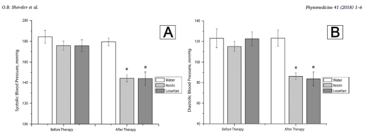
After seven weeks, Reishi therapy reduced blood pressure in experimental animals at a level comparable to that of losartan. Unlike losartan, intragastric Reishi introduction significantly increases cerebral blood flow and affects cerebral cortex metabolic patterns, shifting the balance of inhibitory and excitatory neurotransmitters toward excitation.
Conclusion:
Changes in cerebral blood flow and ratios of neurometabolites suggests Reishi has a potential nootropic effect.
Modulation of gut microbiota and lipid metabolism in rats fed high-fat diets by Ganoderma lucidum triterpenoids
Aijun Tong, et al., Current Research in Food Science 6 (2023) 100427
Abstract
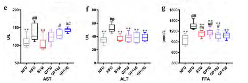
Ganoderma lucidum triterpenoids (GP) have been reported to help prevent and improve hyperlipidemia. Modulation of the gut microbiota was proposed as underlying factor as well as a novel measure to prevent and treat hyperlipidemia. The effects of GP on high-fat diet (HFD)-induced hyperlipidemia and gut microbiota modulation were determined in rats. Ultra-performance liquid chromatography tandem quadrupole time-of-flight mass spectrometry (UPLC-QTOF MS-MS) indicated that GP were enriched with ganoderic acids G, B, H, A, and F. After feeding with GP supplementation, serum lipid levels including total triglyceride, total cholesterol, and low density-lipoprotein cholesterol were significantly decreased in hyperlipidemic rats. Furthermore, administration of GP also has reversed the HFD-induced gut microbiota dysbiosis, including a significant increase in Alloprevotella and reduced proportion of Blautia. The result above suggests that GP would be developed as a functional food to ameliorate lipid metabolic disorders and hyperlipidemia.
Randomized clinical trial of an ethanol extract of Ganoderma lucidum in men with lower urinary tract symptoms.
Noguchi M, et al., Asian Journal of Andrology. 10(5):777-85, 2008.
Abstract
AIM:
To evaluate the safety and efficacy of an extract of Ganoderma lucidum that shows the strongest 5alpha-reductase inhibitory activity among the extracts of 19 edible and medicinal mushrooms by a double-blind, placebo-controlled, randomized and dose-ranging study in men with lower urinary tract symptoms (LUTS).
METHODS:
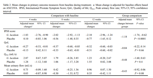
In this trial, we randomly assigned 88 men over the age of 49 years who had slight-to-moderate LUTS to 12 weeks of treatment with G. lucidum extract (6 mg once a day) or placebo. The primary outcome measures were changes in the International Prostate Symptom Score (IPSS) and variables of uroflowmetry. Secondary outcome measures included changes in prostate size, residual urinary volume after voiding, laboratory values and the reported adverse effects.
RESULTS:
G. lucidum was effective and significantly superior to placebo for improving total IPSS with 2.1 points decreasing at the end of treatment (mean difference, -1.18 points; 95% confidence interval, -1.74 to -0.62; P < 0.0001). No changes were observed with respect to quality of life scores, peak urinary flow, mean urinary flow, residual urine, prostate volume, serum prostate-specific antigen or testosterone levels. Overall treatment was well tolerated with no severe adverse effects.
CONCLUSION:
The extract of G. lucidum was well tolerated and improved IPSS scores. These results encouraged a further, large-scale evaluation of phytotherapy for a long duration using the extract of G. lucidum on men with LUTS.
Ganoderma lucidum promotes sleep through a gut microbiota‑dependent and serotonin‑involved pathway in mice.
Chunyan Yao et al., Scientific Reports | (2021) 11:13660
Abstract
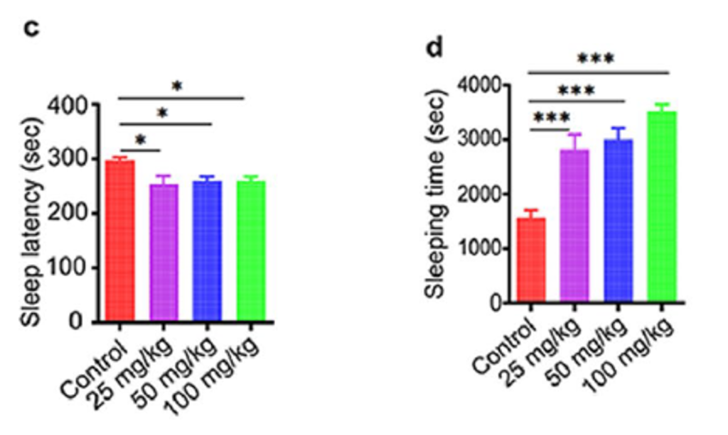
Ganoderma lucidum is a medicinal mushroom used in traditional Chinese medicine with putative tranquilizing effects. However, the component of G. lucidum that promotes sleep has not been clearly identified. Here, the effect and mechanism of the acidic part of the alcohol extract of G. lucidum mycelia (GLAA) on sleep were studied in mice. Administration of 25, 50 and 100 mg/kg GLAA for 28 days promoted sleep in pentobarbital-treated mice by shortening sleep latency and prolonging sleeping time. GLAA administration increased the levels of the sleep-promoting neurotransmitter 5-hydroxytryptamine and the Tph2, Iptr3 and Gng13 transcripts in the sleep-regulating serotonergic synapse pathway in the hypothalamus during this process. Moreover, GLAA administration reduced lipopolysaccharide and raised peptidoglycan levels in serum. GLAA-enriched gut bacteria and metabolites, including Bifidobacterium, Bifidobacterium animalis, indole-3-carboxylic acid and acetylphosphate were negatively correlated with sleep latency and positively correlated with sleeping time and the hypothalamus 5-hydroxytryptamine concentration. Both the GLAA sleep promotion effect andthe altered faecal metabolites correlated with sleep behaviours disappeared after gut microbiota depletion with antibiotics. Our results showed that GLAA promotes sleep through a gut microbiota-dependent and serotonin-associated pathway in mice.
Neurometabolic Effect of Altaian Fungus Ganoderma lucidum (ReishiMushroom) in Rats Under Moderate Alcohol Consumption.
Oleg B. Shevelev, et al., Alcohol Clin Exp Res, Vol 39, No 7, 2015: pp 1128–1136
Background:
we expect neuroprotective prophylactic effects of Reishi-based medications in alcohol-treated animals.
Methods:
The Reishi (R) suspension was produced as water extract from Altaian mushrooms.
Results:
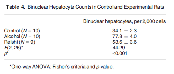
20 days of alcohol consumption significantly increased the number of binuclear hepatocytes compared to the control. This effect was mitigated in the rats that received the Reishi extract.
Conclusions:
Regular administration of the Reishi suspension improved the energy supply to the brain cortex and decreased the prevalence of inhibitory neurotransmitters that are characteristic of alcohol consumption. The alcohol-induced increase in liver proliferation was significantly suppressed by regular administration of the G. lucidum water suspension.
Ganoderic Acids Prevent Renal Ischemia Reperfusion Injury by Inhibiting Inflammation and Apoptosis.
Guangying Shao et al., Int. J. Mol. Sci. 2021, 22, 10229.
Abstract:
Renal ischemia reperfusion injury (RIRI) is one of the main causes of acute kidney injury (AKI), which can lead to acute renal failure. The development of RIRI is so complicated that it involves many factors such as inflammatory response, oxidative stress and cell apoptosis. Ganoderic acids (GAs), as one of the main pharmacological components of Ganoderma lucidum, have been reported to possess anti-inflammatory, antioxidant, and other pharmacological effects. The study is aimed to investigate the protective effect of GAs on RIRI and explore related underlying mechanisms.
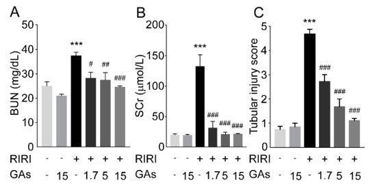
The mechanisms involved were assessed by a mouse RIRI model and a hypoxia/reoxygenation model. Compared with sham-operated group, renal dysfunction and morphological damages were relieved markedly in GAs-pretreatment group. GAs pretreatment could reduce the production of pro-inflammatory factors such as IL-6, COX-2 and iNOS induced by RIRI through inhibiting TLR4/MyD88/NF-kB signaling pathway. Furthermore, GAs reduced cell apoptosis via the decrease of the ratios of cleaved caspase-8 and cleaved caspase-3. The experimental results suggest that GAs prevent RIRI by alleviating tissue inflammation and apoptosis and might be developed as a candidate drug for preventing RIRI-induced AKI.
Ganoderic Acid A Inhibits Bleomycin-Induced Lung Fibrosis in Mice.
Gaoyan Wen et al., Pharmacology DOI: 10.1159/000505297, 2019
Abstract
Background:
To study the protective effects of ganoderic acid A (GAA) on bleomycin (BLM)-induced pulmonary fibrosis.
Methods:
ICR mice were intratracheally instilled with BLM to induce pulmonary fibrosis on day 0. Then the mice were orally given GAA (25, 50 mg/kg) or dexamethasone (2 mg/kg). After treatment for 21 days, the mice were sacrificed. Wet dry weight (W/D) ratio of lung was used to detect pulmonary edema. Myeloperoxidase (MPO), interleukin-1β (IL-1β), IL-6, tumor necrosis factor-α (TNF-α), malondialdehyde (MDA), and superoxide dismutase (SOD) were detected by enzyme-linked immunosorbent assay. Hematoxylin and eosin staining was used to evaluate the pathological changes. The levels of transforming growth factor β (TGF-β), phosphorylated-smad3 (p-smad3), p-IκB, and p-nuclear factor kappa B (NF-κB) in lung tissue were detected by western blot.
Results:
GAA treatment significantly improved MPO activity, W/D ratio, and lung histopathology. The protective effect of GAA may be related to downregulation of TNF-α, IL-1β, IL-6, MDA and upregulation of SOD. In addition, GAA significantly decreased the levels of TGF-β, p-smad3, p-IκB, and p-NF-κB, compared with those in BLM group.
Conclusion:
GAA has protective effect on BLM-induced lung injury, and TGF-β/Smad-3/NF-κB signaling pathway may play an important role in the pathogenesis of BLM-induced lung injury.

Results of long-term carcinogenicity bioassays on Coca-Cola administered to Sprague-Dawley rats.
Belpoggi F, et al., Annals of the New York Academy of Sciences. 1076:736-52, 2006.
Abstract:
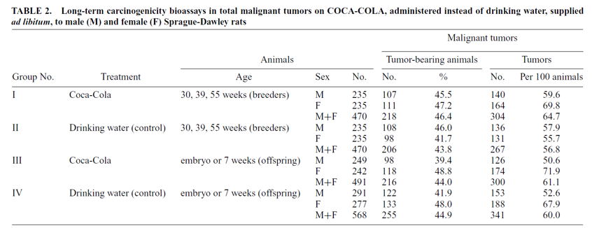
Coca-Cola is currently sold in more than 200 countries and in early 2000, the company sold its 10 billionth unit case of Coca-Cola branded products. Given the worldwide consumption of Coca-Cola, a project of experimental bioassays to study its long-term effects when administered as substitute for drinking water on male and female Sprague-Dawley rats was planned and executed. The objective of the project was to study whether and how long-term consumption of Coca-Cola affects the basic tumorigram of test animals. The bioassays were performed on rats beginning at different ages, namely: (a) on males and females exposed since embryonic life or from 7 weeks of age; and (b) on males and females exposed from 30, 39, or 55 weeks of age. Overall, the project included 1999 rats. During the biophase, data were collected on fluid and feed consumption, body weight, and survival. Animals were kept under observation until spontaneous death and underwent complete necropsy. The results indicate: (a) an increase in body weight in all treated animals; (b) a statistically significant increase of the incidence in females, both breeders and offspring, bearing malignant mammary tumors; (c) a statistically significant increase in the incidence of exocrine ademonas of the pancreas in both male and female breeders and offspring; and (d) an increased incidence, albeit not statistically significant, of pancreatic islet cell carcinomas in females, a malignant tumor which occurs very rarely in our historical controls. On the basis of the results of this study, excessive consumption of regular soft-drinks should be generally discouraged, in particular for children and adolescents.
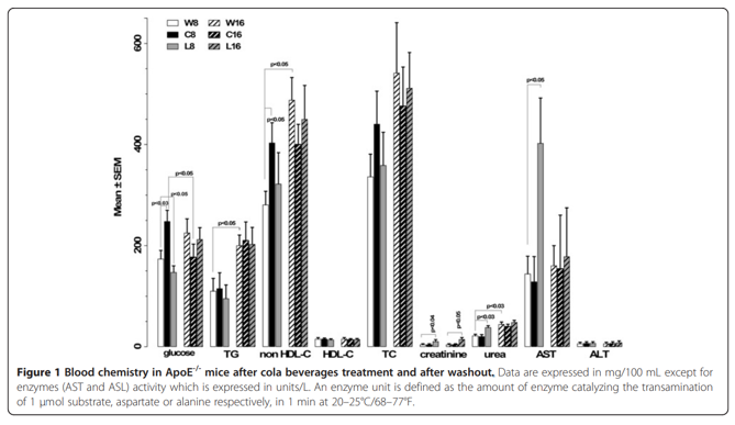
Metabolic disturbances and worsening of atherosclerotic lesions in ApoE-/- mice after cola beverages drinking.
Otero-Losada ME, et al., Cardiovascular Diabetology. 12:57, 2013.
BACKGROUND:
Atherosclerosis is a major health burden. Metabolic disorders had been associated with large consumption of soft drinks. The rising incidence of atherosclerosis and metabolic alterations warrants the study of long-term soft drink consumption’ effects on metabolism and atherosclerosis in genetic deficiency of apolipoprotein E which typically develops spontaneous atherosclerosis and metabolic alterations.
METHODS:
ApoE-/- mice were randomized in 3 groups accordingly with free access to: water (W), regular cola (C) or light cola (L). After 8 weeks, 50% of the animals in each group were euthanized.
CONCLUSION:
Cola beverages caused atherosclerotic lesions’ enlargement with metabolic (C) or non metabolic disturbances (L). ApoE-/- mice were particularly sensitive to L treatment. These findings may likely relate to caramel colorant and non-nutritive sweeteners in cola drinks and have potential implications in particularly sensitive individuals.
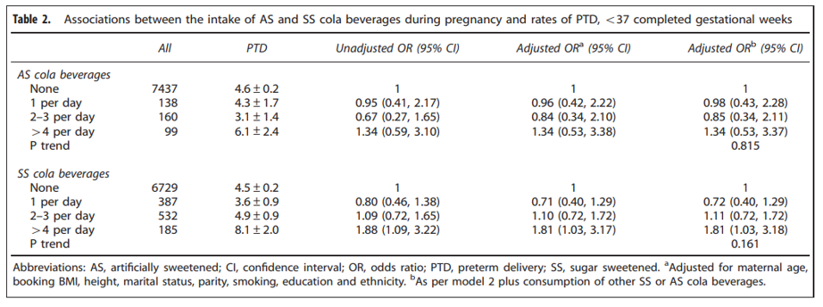
Relationship between artificially sweetened and sugar-sweetened cola beverage consumption during pregnancy and preterm delivery in a multi-ethnic cohort: analysis of the Born in Bradford cohort study.
Petherick ES, et al., European Journal of Clinical Nutrition. 68(3):404-7, 2014.
Abstract:
The aim of this study was to investigate the relationship between the intake of sugar-sweetened (SS) and artificially sweetened (AS) cola beverages during pregnancy and the risk of preterm delivery (PTD). At baseline (2007-2010), 8914 pregnant women were recruited to the Born in Bradford birth cohort study at 24-28 weeks of pregnancy. Women completed a questionnaire describing their health and lifestyle behaviours, including their consumption of AS and SS cola beverages reported as cups per day, which were then linked to maternity records. The relationship between SS and AS cola beverage consumption was examined using logistic regression analyses. No relationship was observed between daily AS cola beverage consumption and PTD. Women who drank four cups per day of SS cola beverages had higher odds of a PTD when compared with women who did not consume these beverages daily. We conclude that high daily consumption of SS cola beverages during pregnancy is associated with increases in the rate of PTD.
Cardiorenal Involvement in Metabolic Syndrome Induced by Cola Drinking in Rats: Proinflammatory Cytokines and Impaired Antioxidative Protection.
Otero-Losada M, Mediators of Inflammation. 561: 3056, 2016.
Abstract:
We report experimental evidence confirming renal histopathology, proinflammatory mediators, and oxidative metabolism induced by cola drinking. Male Wistar rats drank ad libitum regular cola (C, n = 12) or tap water (W, n = 12). Measures. Body weight, nutritional data, plasma glucose, cholesterol fractions, TG, urea, creatinine, coenzyme Q10, SBP, and echocardiograms (0 mo and 6 mo). Chronic cola drinking induced cardiac remodeling associated with increase in proinflammatory cytokines and renal damage. Hypertriglyceridemia and oxidative stress were key factors. Hypertriglyceridemic lipotoxicity in the context of defective antioxidant/anti-inflammatory protection due to low Q10 level might a key role in cardiorenal disorder induced by chronic cola drinking in rats.

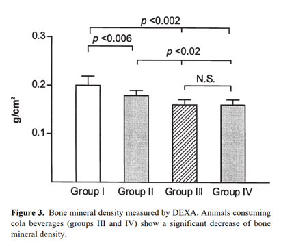
Cola beverage consumption induces bone mineralization reduction in ovariectomized rats.
Garcia-Contreras F, et al., Archives of Medical Research. 31(4):360-5, 2000 Jul-Aug.
BACKGROUND:
A significant association of cola beverage consumption and increased risk of bone fractures has been recently reported. The present study was carried out to examine the relationship of cola soft drink intake and bone mineral density in ovariectomized rats.
METHODS:
Study 1. Four groups of 10 female Sprague-Dawley rats were studied. Animals from groups II, III, and IV were bilaterally ovariectomized. Animals from groups I and II received tap water for drinking, while animals from groups III and IV each drank a different commercial brand of cola soft drink. After 2 months on these diets, the following were measured: solid diet and liquid consumption; bone mineral density; calcium in bone ashes; femoral cortex width; calcium; phosphate; albumin; creatinine; alkaline phosphatase; 25-OH hydroxyvitamin D, and PTH.
CONCLUSIONS:
These data suggest that heavy intake of cola soft drinks has the potential of reducing femoral mineral density.
